Itchy Scrotum: Is It Eczema, Fungal Infection, or Something Else?
Scrotal pruritus remains significantly under-reported relative to its actual prevalence in adult male dermatology. Symptom severity varies considerably — from intermittent low-intensity irritation to nocturnal itch of sufficient magnitude to fragment sleep architecture and impair occupational functioning.
Scrotal eczema, jock itch, candidal intertrigo, and contact dermatitis all generate erythema, pruritus, and cutaneous disruption within the same anatomical zone — yet the treatment algorithm for each condition differs fundamentally. Misidentification does not merely postpone clinical improvement; inappropriate therapy actively exacerbates the underlying pathology.
From a prognostic standpoint, scrotal pruritus is overwhelmingly benign. The clinical imperative lies not in excluding serious disease — which is rarely the cause — but in arriving at a precise aetiological diagnosis that directs appropriate treatment.
Common Causes of an Itchy Scrotum
Scrotal skin is a physiological outlier. Permeability studies estimate it absorbs topical substances at rates 40 to 80 times higher than forearm skin (1). That degree of absorptive capacity makes the scrotum disproportionately reactive to irritants, allergens, heat, moisture, and mechanical friction — which explains why so many distinct pathologies cluster here and present with frustratingly similar morphology on examination.
The differential for itchy balls causes and groin irritation spans several categories. Scrotal eczema — technically scrotal dermatitis — predominates among chronic presentations. Tinea cruris, the dermatophyte infection commonly termed jock itch, constitutes the principal infectious aetiology. Candidal overgrowth — the genital yeast infection that clinicians classify as candidal intertrigo — preferentially colonises occluded intertriginous skin under conditions of sustained warmth and humidity. Irritant and allergic contact dermatitis are underdiagnosed contributors frequently misclassified as “eczema” without further workup. STIs — herpes simplex, condylomata, pediculosis pubis — are occasionally implicated, though virtually never as the sole explanation for scrotal pruritus absent other genital findings. Mechanical irritation secondary to synthetic textiles or poorly fitted undergarments is another contributor that clinical assessments frequently underweight.
Misdiagnosis carries real clinical consequences. Applying an antifungal to eczematous scrotal skin accomplishes nothing. A potent topical steroid applied to an active dermatophyte infection suppresses visible inflammation while the fungus proliferates beneath (2).
What Is Scrotal Eczema?
Scrotal dermatitis — the more precise designation for scrotal eczema — represents a chronic inflammatory dermatosis localised to the thin, hyperabsorptive scrotal epithelium. No bacterium, fungus, or virus drives it. Pathophysiologically, the condition reflects aberrant immune activation: cutaneous inflammatory cascades trigger against ordinarily innocuous stimuli. Disease trajectory follows a relapsing-remitting pattern, with episodes recurring over months to years at fluctuating severity (1).
Three clinical subtypes constitute the majority of diagnosed cases. Atopic dermatitis is the most frequently encountered variant, with a well-documented hereditary basis, patients carrying active lesions on facial skin, the antecubital fossae, or popliteal fossae demonstrate markedly elevated incidence of concurrent scrotal disease. Contact dermatitis, both irritant and allergic, is triggered by direct substance exposure: fragranced wash products, latex condoms, laundry detergent residues, spermicidal agents, or paradoxically, topical medications applied to treat an earlier flare. Seborrhoeic dermatitis occasionally involves the scrotum, presenting with flaky, somewhat greasy scales (3).
Severity ranges from mild well-demarcated erythema that clears upon irritant removal, through moderate diffuse redness extending onto the penile shaft or inguinal folds, to severe presentations with weeping, erosions, discharge, and secondary bacterial superinfection risk — at which point formal dermatological assessment becomes necessary (3).
One distinction that warrants explicit clarification, given how frequently patients raise the concern: scrotal eczema has no transmissible component. Neither sexual nor incidental skin-to-skin contact poses any risk of spread — a fundamentally different profile from the infectious aetiologies discussed below.
Eczema vs. Fungal Infection vs. Other Conditions
This is where jock itch vs eczema confusion most commonly leads to weeks of misdirected self-treatment.
Anatomical distribution is the single most useful clinical discriminator. Tinea cruris, caused predominantly by Trichophyton rubrum, begins in the crural fold and fans outward across the medial thighs. The scrotum itself is typically spared or only marginally involved (2, 5). When scrotal skin is the primary site of inflammation, dermatophyte infection drops down the differential and eczema or candidal intertrigo becomes more probable.
Morphology reinforces the distinction. A jock itch rash has a textbook annular configuration: a raised, scaly advancing border with relative central clearing (5, 6). A rash on scrotum driven by eczema presents as diffuse, poorly demarcated erythema with surface dryness and, in chronic cases, lichenification — but no ring-shaped border (1, 3).
Candida albicans intertrigo produces beefy red, slightly macerated rash in occluded skin folds with satellite pustules scattered just beyond the rash margin — a nearly pathognomonic finding. Patients with diabetes, elevated BMI, or recent antibiotic exposure are disproportionately affected (4).
Sexually transmitted infections rarely manifest as isolated scrotal pruritus. Herpes simplex produces grouped vesicles or painful shallow erosions; condylomata acuminata present as verrucous papules; Phthirus pubis causes visible excoriations and nits. Without these additional features, an STI is a statistically unlikely explanation — though testing remains appropriate when clinical doubt exists.
When clinical ambiguity persists, a potassium hydroxide (KOH) preparation from a skin scraping resolves the question within minutes (2).
Symptoms and Triggers
The dominant complaint is pruritus — intense itch that drives the majority of clinical presentations. Accompanying signs include erythema, surface dryness, fine scaling, and in long-standing cases, lichenification from chronic scratching. Contact dermatitis variants frequently add a burning component. More active disease may produce serous weeping and superficial erosions (1, 3).
Trigger identification is therapeutically essential. Fragranced soaps and shower gels, scented laundry detergents depositing chemical residue onto underwear fabric, synthetic underwear materials trapping heat and moisture, latex condoms and spermicidal products, excessive perspiration and friction from athletic activity, and psychological stress — a well-established precipitant of inflammatory dermatoses — all feature prominently as documented exacerbating factors (3, 4).
A practical recommendation is to maintain a brief symptom diary over two to three weeks documenting wash products, detergents, underwear fabric, activity levels, stress, and itch episodes. The emerging pattern typically narrows the trigger list far more efficiently than empiric guesswork.
Treatment and Relief
Effective management operates on two parallel tracks: dampening active inflammation and restructuring the skin environment to reduce flare frequency.
Fragrance-free emollients are the non-negotiable foundation — they reconstitute the compromised epidermal barrier, counteract transepidermal water loss, and materially reduce flare frequency when applied consistently, particularly within minutes of bathing (3). Gel-based formulations tend to be better tolerated on scrotal skin than heavy ointments. Emollients should replace soap entirely in the affected area.
For active flares, mild-to-moderate potency topical corticosteroids remain first-line, though genital skin’s amplified absorption means a mid-potency steroid delivers pharmacological effects equivalent to a much stronger preparation used elsewhere. Duration should be limited and clinician-supervised (3). For maintenance therapy, topical calcineurin inhibitors — tacrolimus ointment, pimecrolimus cream — carry no atrophy risk and are specifically indicated for sensitive anatomical sites (7).
When eczema on penis accompanies scrotal dermatitis, the therapeutic framework remains consistent, though penile skin’s heightened steroid absorption requires specialist-level oversight.
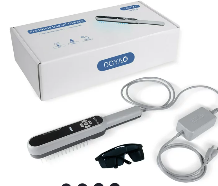
Moderate-to-severe scrotal eczema that fails to achieve adequate control with topical therapy alone may respond to narrow-band UVB phototherapy, which has yielded statistically significant improvements in both subjective pruritus scores and objective inflammatory measures. A thorough clinical discussion of this modality appears in the UVB light therapy section of this site.
Multi-site disease — particularly when facial involvement coexists — benefits from a unified management strategy. The treatment paradigm for eczema on face operates on identical foundational principles, though periorbital and perioral anatomy necessitate distinct therapeutic modifications.
Confirmed tinea cruris requires an entirely separate therapeutic pathway. Topical antifungals — clotrimazole, miconazole, terbinafine, or ketoconazole — administered twice daily over 10 to 14 days achieve mycological cure in approximately 80 to 90 percent of uncomplicated infections (2, 6). Coexistent tinea pedis functions as a dermatophyte reservoir capable of reseeding the inguinal region and demands concurrent treatment. A broader examination of inflammatory versus infectious scrotal pathology appears in the laser treatments for eczema article. For a consolidated reference across the full severity spectrum, the eczema treatment guide provides comprehensive coverage.
When to See a Doctor
Uncomplicated mild presentations are appropriate candidates for an initial conservative trial: withdrawal of fragranced products, initiation of a fragrance-free emollient, transition to non-occlusive cotton undergarments, and a two- to three-week reassessment interval.
Referral for formal evaluation is indicated when symptoms persist beyond the trial period without measurable improvement, when lesion morphology evolves or extends, when vesiculation, erosion, or purulent exudate develops, when clinical features suggest secondary bacterial superinfection, or when diagnostic ambiguity persists.
Dermatological consultation enables diagnostic confirmation, KOH microscopy for definitive mycological assessment, epicutaneous patch testing to unmask occult contact sensitisers, and histopathological examination of ambiguous lesions. Establishing an accurate diagnosis early compresses the treatment timeline and forestalls progressive lichenification and secondary infection (1).
Pruritus sine materia — itch in the absence of visible cutaneous pathology — is a well-documented presentation in scrotal dermatology. Subclinical eczema, pronounced xerosis, and subthreshold allergic contact sensitisation all produce pruritus prior to overt dermatitis. A neurogenic aetiology has also been characterised in a subset of patients: peripheral or central nociceptive pathways amplify itch transmission independently of identifiable skin disease, partially accounting for inadequate responses to conventional topical therapies (7). Itch persisting beyond two weeks without discernible precipitant warrants formal dermatological assessment.
Anatomically: dermatophytes originate in the inguinal fold and track along inner thigh skin while sparing the scrotum. Eczema does the opposite — scrotal skin is primary. Morphologically: tinea forms annular plaques with a raised scaly advancing rim; eczema produces diffuse erythema, indistinct borders, no ring pattern (2, 5). KOH microscopy resolves diagnostic uncertainty within minutes
Irritant contact dermatitis may clear within weeks once the offending agent is withdrawn. Atopic scrotal eczema follows a chronic relapsing course requiring sustained barrier maintenance. Untreated tinea cruris rarely self-clears and risks dermatophyte dissemination to toenails and interdigital spaces (2, 6).
Isolated scrotal pruritus absent vesicles, erosions, verrucous growths, or urethral discharge carries a very low pre-test probability for any STI. Eczema, tinea cruris, and contact dermatitis explain the vast majority of presentations seen in dermatology clinics. That said — even a single atypical feature or a relevant exposure history should prompt panel testing without delay
Accuracy determines speed. For eczema: withdrawing fragranced products and initiating emollient therapy with a low-potency corticosteroid yields perceptible pruritus reduction within 72 to 120 hours. For dermatophytosis: twice-daily topical antifungal application produces visible improvement by day five to seven (2). Both benefit from concurrent switch to non-occlusive cotton undergarments. Persistent diagnostic uncertainty is best resolved through a single dermatology consultation
To provide the best experiences, we use technologies like cookies to store and/or access device information. Consenting to these technologies will allow us to process data such as browsing behavior or unique IDs on this site. Not consenting or withdrawing consent, may adversely affect certain features and functions.
Functional
Always active
The technical storage or access is strictly necessary for the legitimate purpose of enabling the use of a specific service explicitly requested by the subscriber or user, or for the sole purpose of carrying out the transmission of a communication over an electronic communications network.
Preferences
The technical storage or access is necessary for the legitimate purpose of storing preferences that are not requested by the subscriber or user.
Statistics
The technical storage or access that is used exclusively for statistical purposes.The technical storage or access that is used exclusively for anonymous statistical purposes. Without a subpoena, voluntary compliance on the part of your Internet Service Provider, or additional records from a third party, information stored or retrieved for this purpose alone cannot usually be used to identify you.
Marketing
The technical storage or access is required to create user profiles to send advertising, or to track the user on a website or across several websites for similar marketing purposes.