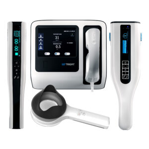
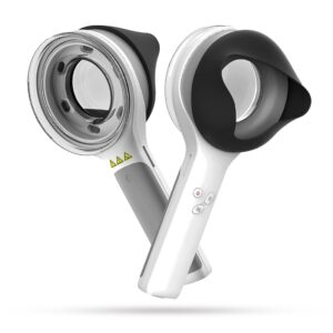
Wood Lamps KERNEL for Skin Analysis


Info
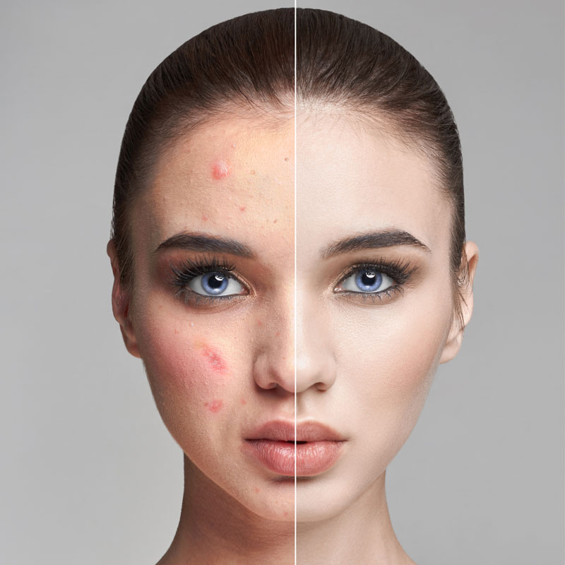
Hardly any diagnostic instrument in skin care has stuck around the way the Wood lamp has. Aim its long-wave ultraviolet beam at the skin in a darkened room, and details that ordinary lighting keeps hidden begin to surface: a fading pigment border here, an infected patch there. Clinicians have relied on the Wood lamp for skin analysis since the early 1900s, and it still sees daily use across dermatology and aesthetic consultations (1). UVTREAT supplies clinic-grade Wood’s lamps KERNEL and other professional skin analysis equipment to dermatology and aesthetic practices throughout the U.S. and Latin America.
How Wood Lamp Skin Examination Works in Clinical Practice
Its output sits in the 320–400 nm band, peaks at about 365 nm, and a filter inside the head strips out almost all visible light (2). UV doesn’t register to the eye, so the head itself glows a dim violet, and unaffected skin comes back as a calm, even blue with no fluorescence. Trouble shows up as a change. An area that fluoresces, brightens, or shifts color tells the examiner something is there.
Running the test takes little effort. Black out the room, give the lamp a minute or so to warm up, and bring it within 10 to 30 cm of the skin once the eyes have adjusted (3). Nothing about it hurts, and the head stays cool. The one instruction patients tend to forget: come with clean skin that hasn’t just been washed, and skip makeup, lotion, and deodorant, all of which fluoresce by themselves and throw the reading off (2). Interpreting what appears is the harder part, and it rewards experience.
Most of the diagnostic value comes down to wood’s lamp color diagnosis, since individual conditions tend to fluoresce in recognizable shades. Take erythrasma, a surface bacterial infection caused by Corynebacterium minutissimum: the coproporphyrin it secretes lights up an unmistakable coral-red (4). Pseudomonas reads green. Of the dermatophytes, the Microsporum species responsible for much of tinea capitis (scalp ringworm) glow blue-green, while tinea versicolor tends toward yellow-green (2). Oily zones look yellow, thickened patches white, and stray fibers or lint usually flash bright white too.
Pigment work is the other major use. During wood lamp for vitiligo assessment, depigmented patches turn a sharp, bright blue-white with crisp edges, which makes faint or early lesions far easier to outline than they are under room lighting (4). That same contrast lets clinicians track disease stability and watch repigmentation as treatment progresses. Newer narrowband 365-nm lamps push this contrast further still, outperforming the older broadband designs for pigmentary cases (5). When pigment loss needs confirmation at the cellular level, some practitioners pair the lamp with reflectance confocal microscopy to guide vitiligo staging (6). Epidermal melasma and other hyperpigmentation stand out more clearly under the lamp as well.
In aesthetic and cosmetology work, a wood light skin examination tends to come first, before any facial, peel, or light-based procedure. The practitioner notes oiliness, dehydration, how oxidized the comedones are, any scaling, and how deeply the pigment sits, then shapes the protocol from there. A dermatology clinic, an aesthetic center, a cosmetology studio – wherever it happens, the wood lamp Kernel skin examination adds quick, hands-off detail to what the patient’s history already shows. Worth keeping in mind, though: it reads conditions rather than treating them, and since many fungi and bacteria never fluoresce, a clean result on its own doesn’t clear the patient (3).
Choosing a Professional Wood Lamp KERNEL
Performance varies a great deal between units. UV consistency is the first thing to scrutinize. A stable, properly filtered source around 365 nm gives dependable fluorescence, and as noted, narrowband lamps deliver noticeably sharper contrast for pigment work than broadband ones (5). After that, look at construction. Medical-grade housing survives daily handling, and a sensible grip keeps the hand from tiring across a full clinic day.
Portability earns its keep in practices that rotate between rooms or run assessments off-site, and enough raw intensity matters when a treatment room won’t go fully dark. The device also has to slot into real workflow, with quick warm-up and simple handling that add seconds to a visit rather than minutes. UVTREAT designs its professional Wood lamp Kernel line around exactly this kind of dermatology and aesthetic use, and it sits within a broader phototherapy catalogue that includes UV lamps for psoriasis treatment.
Final Thoughts
The Wood lamp has stayed in everyday use for a simple reason: it is quick, inexpensive, and tells the specialist something real. In seconds it can outline vitiligo, raise a flag on fungal or bacterial infection, and tighten up a pigment assessment. What turns those quick readings into something a clinician can act on with confidence is dependable, clinic-grade hardware.
References
- Oakley A. Wood lamp skin examination. DermNet NZ. Available at: https://dermnetnz.org/topics/wood-lamp-skin-examination
- Al Aboud DM, Gossman W. Wood’s Light. StatPearls. Treasure Island (FL): StatPearls Publishing. Available at: https://www.ncbi.nlm.nih.gov/books/NBK537193/
- Cleveland Clinic. Wood’s Lamp Examination. Available at: https://my.clevelandclinic.org/health/diagnostics/23292-woods-lamp-examination
- Dyer JM, Foy VM. Revealing the Unseen: A Review of Wood’s Lamp in Dermatology. J Clin Aesthet Dermatol. 2022;15(6):25–30. Available at: https://jcadonline.com/review-of-woods-lamp-in-dermatology/
- Bae JM, et al. A 365-nm narrowband Wood’s lamp for vitiligo and hypopigmentation disorders. J Am Acad Dermatol. Available at: https://www.jaad.org/article/S0190-9622(19)32670-2/abstract
- Hou Y, et al. In-depth study of Wood’s lamp examination combined with reflective confocal laser scanning microscopy for the guidance of vitiligo staging and treatment. J Cosmet Dermatol. 2024;23(4):1472–1479. Available at: https://onlinelibrary.wiley.com/doi/10.1111/jocd.16145
FAQ
- Dermatologists shine it on skin and scalp to surface what daylight misses - pigment disorders, fungal or bacterial infection, and various surface irregularities. It feeds into diagnosis and planning, but the lamp assesses; it doesn't treat.
- With the room dark and the lamp warmed up, the clinician brings it within a few inches of clean skin. Normal skin stays dark and bluish; problem areas shift color or fluoresce, which hints at what lies beneath.
- It can. Under the lamp, vitiligo patches go bright blue-white with crisp edges, so faint or early lesions become easier to find, map, and follow over time - especially on lighter skin.
- Each shade hints at a cause. Coral-red leans toward erythrasma, green toward Pseudomonas, blue-green toward some ringworm fungi, yellow-green toward tinea versicolor, and bright blue-white toward pigment loss like vitiligo. On healthy skin, nothing fluoresces.