HomeArticlesWhat Is a Colposcope? A Complete Guide for Modern Gynecology
Written byAnna Chacon,MD; Board-Certified Dermatologist
Published on July 14, 2026
What Is a Colposcope? A Complete Guide for Modern Gynecology
Cervical examination remains a fundamental component of preventive gynecological care. The overwhelming majority of cervical cancers develop from persistent infection with high-risk human papillomavirus (HPV), progressing through histologically detectable precancerous stages over an extended interval (1)(2). This prolonged preinvasive phase accounts for the diagnostic value of screening and magnified visual assessment, since intraepithelial abnormalities can be identified and managed well before invasion occurs. The instrument that enables sufficiently detailed inspection of the cervix to detect such early changes is the colposcope.
In clinical practice, colposcopy is indicated when a screening result warrants further evaluation of the cervix. The procedure does not substitute for screening, nor does it establish a definitive diagnosis in isolation; rather, it occupies the interval between an abnormal screening result and histological confirmation. This article addresses the definition and function of the colposcope, its operating principle, its clinical applications, the difference between colposcopy and biopsy, the principal device categories, and the criteria relevant to equipment selection.
What Is a Colposcope?
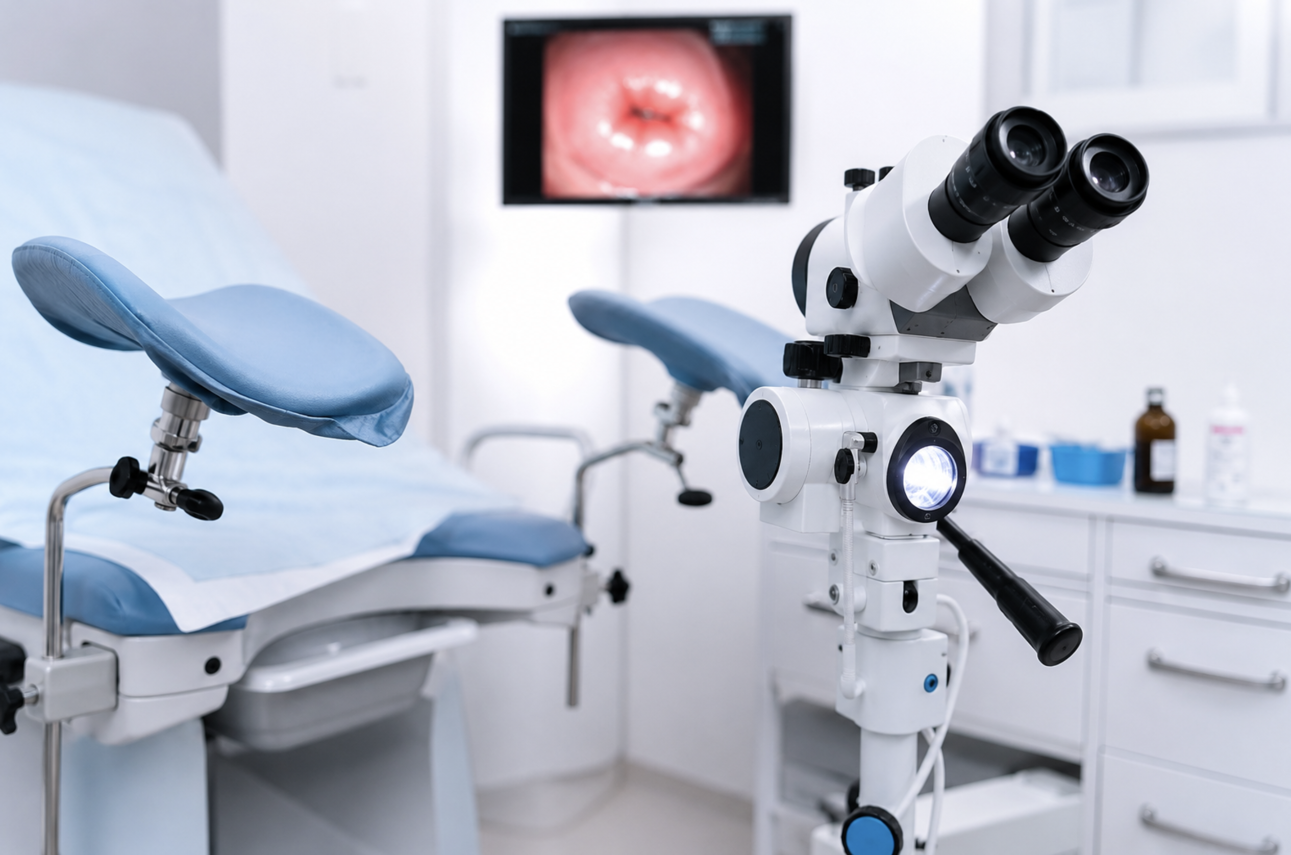
A colposcope is a low-power binocular magnifying instrument coupled with a high-intensity light source, employed to examine the cervix, vagina, and vulva under illumination across a range of magnifications (3). The International Agency for Research on Cancer characterizes it as a comparatively simple device comprising a binocular microscope and a light source, frequently fitted with a beam splitter to permit attachment of a still or video camera (3). The instrument does not contact the patient; it magnifies and illuminates the tissue from a fixed working distance (4).
The main components are consistent across models. A binocular (stereoscopic) head with two independently focusable eyepieces affords the depth perception required for directed biopsy and excisional treatment (3). An objective lens with a focal length of approximately 300 mm establishes the working distance, proximate enough to access the cervix yet sufficient to accommodate the passage of instruments (3)(5). A magnification changer, an illumination source (halogen or LED), and a green (red-free) filter constitute the remainder of the optical system, and most contemporary units incorporate a camera port for documentation (3).
Hans Hinselmann introduced colposcopy in Germany in 1925, and it has been a standard way of working up abnormal cytology ever since (6). What sets it apart from a routine gynecological exam is straightforward. In a standard exam the speculum simply opens the vaginal walls so the cervix can be seen. The colposcope adds magnification, a strong standardized light, and filtered viewing that bring out epithelial and vascular changes the naked eye would miss – and it lets the clinician place a biopsy precisely (3)(4). The range of available colposcopes follows from this core function
How Does a Colposcope Work?
A colposcope functions by magnifying and intensely illuminating cervical tissue, rendering surface architecture and vascular patterns visible. Lower magnifications of approximately 2× to 6× provide an overview of surface architecture, whereas higher settings, typically ranging from 8× to 15× and reaching 25× to 30× on certain models, serve to examine fine detail and vascular patterns (7). In clinical use, magnification exceeding 15× is seldom required, and three to four settings between 4× and 15× are generally adequate (3). Increasing magnification narrows the field of view and diminishes the depth of focus; greater magnification is therefore not invariably advantageous.
Illumination and optics are equally important. Light sources are either halogen, which is powerful, inexpensive, and easily replaced, or LED, which lasts considerably longer (3). The green (red-free) filter is a defining feature: by removing background red, it makes blood vessels appear dark, which sharpens the detection of vascular patterns such as punctation and mosaicism (3). In digital and video systems, a camera and monitor are added, allowing images to be captured, displayed, and stored for later reference (3).
Colposcopy itself is a stepwise, dynamic examination. The patient is positioned in a semi-lithotomy position and a speculum is inserted so the entire cervix can be viewed at low power (3)(6). Normal saline may be applied first to study the subepithelial vasculature with the green filter. Then 3% to 5% acetic acid is applied liberally. Acetic acid clears mucus and reacts with nuclear proteins, causing areas of increased nuclear content, such as dysplasia, to turn white, an effect known as acetowhitening (8). Because this effect fades, acetic acid must be reapplied roughly every few minutes (8). Lugol’s iodine may then be applied – the basis of the Schiller’s test. Normal squamous epithelium, being rich in glycogen, takes up the stain and turns brown to black; abnormal, glycogen-depleted areas stay pale (3)(9). An iodine-positive area is almost always normal, though the reverse does not hold: not every iodine-negative area is pathological (3).
Throughout the examination, the clinician evaluates the transformation zone and the squamocolumnar junction, assessing the lesion borders, opacity, and vascular features that differentiate low-grade from high-grade changes (3)(6). This process of understanding how colposcopy works underlines an important point: abnormal vascular features are meaningful only within acetowhite areas, and no single feature is uniquely diagnostic (3).
What Is a Colposcope Used For?
A colposcope is used to investigate abnormalities detected during cervical screening and to evaluate suspicious findings elsewhere in the lower genital tract. The most frequent indications are an abnormal Pap (cytology) result and a positive high-risk HPV test (4)(6). A clinically suspicious cervix warrants colposcopy even in the presence of negative screening, as the visual examination may detect changes that testing does not (6).
Beyond initial evaluation, colposcopy is central to assessing and monitoring cervical intraepithelial neoplasia (CIN), graded 1 through 3, along with other HPV-related lesions (4)(6). Its use extends to the vagina and vulva as well: vaginal intraepithelial neoplasia (VaIN) and vulvar intraepithelial neoplasia (VIN) are evaluated colposcopically, which matters because HPV tends to affect the lower genital tract as a field, and lesions in the vaginal vault are easily overlooked (6). Colposcopy is also used for surveillance after treatment of high-grade disease (6).
Modern management frameworks have refined when colposcopy is recommended. Under the risk-based approach adopted by the American Society for Colposcopy and Cervical Pathology, the decision to refer depends on a patient’s calculated risk of CIN 3 or worse rather than on a single test result (10). Colposcopy is recommended when the immediate risk of CIN 3+ is at least 4% but below 25%; at higher risk thresholds, treatment may be considered without a preceding biopsy (10)(11). Accordingly, colposcopy constitutes a defined step within the screening pathway rather than a routine response to every abnormal result.
Colposcopy vs Biopsy: What Is the Difference?
The distinction between colposcopy and biopsy is frequently misunderstood by patients and is clinically significant. Colposcopy is a visual diagnostic examination that localizes and characterizes abnormal areas but yields an impression rather than a tissue diagnosis. A biopsy removes a small tissue sample for histopathological examination, and histology remains the diagnostic gold standard for CIN and cervical cancer (12).
In practice, the two are combined as colposcopy-directed biopsy, in which the colposcope identifies the most abnormal area and guides the site of sampling (12). Neither procedure substitutes for the other. Colposcopy performed without biopsy provides only a subjective, operator-dependent impression, whereas a biopsy obtained without colposcopic guidance carries a substantial risk of sampling error.
The accuracy data explain why both are needed. The sensitivity of a colposcopic impression for detecting CIN 2+ varies considerably according to the threshold applied, with a reported weighted mean sensitivity of approximately 68.5% and specificity of approximately 75.9% under one common definition (13). Sampling strategy is likewise consequential: a single biopsy of the most abnormal-appearing area may fail to detect up to one-third of precancers, and sensitivity increases substantially as additional targeted samples are obtained. For this reason, current standards recommend obtaining at least two, and up to four, targeted biopsies of distinct abnormal areas (12)(14). The visual examination and the tissue sample are two halves of a single diagnostic process.
Types of Colposcopes: Digital vs Optical Systems
Colposcopes are classified into two principal categories, optical and digital, with portable devices representing an expanding third group. Each category presents distinct advantages, and the appropriate selection depends substantially on the clinical setting.
Optical (traditional binocular) colposcopes provide direct stereoscopic viewing through eyepieces. This confers excellent depth perception and true three-dimensional visualization, which is advantageous for directed biopsy and excisional procedures, and the instruments are durable and well established (3). Their limitations are ergonomic and practical: the examiner works in close proximity to the patient and refocuses manually, and image capture requires a supplementary camera. Notably, comparative work has found that clinicians often rate optical systems as easier for visualization and sampling, with overall clinical outcomes similar between optical and video systems, which tempers any assumption that digital is automatically superior (15).
Digital (video) colposcopes replace the eyepieces with a monitor and use a camera to capture still images and video. Their advantages lie in documentation, integration with electronic medical records, telemedicine, teaching, and patient communication, along with reduced physical strain on the examiner (3). The trade-offs are a reliance on software and storage and, in standard configurations, the loss of true stereoscopic depth. The comparison below summarizes the practical differences.
Feature
Optical (binocular)
Digital (video)
Viewing method
Direct through eyepieces
Monitor display
Depth perception
True stereoscopic (3D)
Typically two-dimensional
Documentation
Requires added camera
Built-in image and video capture
EMR / telemedicine
Limited
Well suited
Teaching and patient communication
Limited
Strong
Examiner ergonomics
Closer working position
Reduced strain
A third category, portable and handheld devices, including smartphone-based systems, extends colposcopy to resource-limited settings and screening programs where standard equipment is unavailable (16). These typically use LED illumination and an integrated green filter in a lightweight form. Optical colposcopy remains well suited to an experienced clinician operating within a straightforward workflow; digital systems are appropriate for practices requiring documentation and teaching capacity; and portable units serve outreach and community-based programs.
How to Choose the Right Colposcope for Your Practice
Selecting a professional colposcope requires matching the device to its intended application. Several factors warrant careful consideration:
Optical quality and magnification. Binocular, stereoscopic viewing is considered essential, and while devices are often advertised across a 3× to 30× range, the practically useful clinical range is roughly 4× to 15× (3).
Illumination. LED sources are generally preferred for their longer life and stable color temperature, and a dedicated green filter for vascular assessment is important (3).
Working distance. A focal length of about 300 mm balances access and reach (3)(5).
Camera and documentation. Image resolution, archiving, and integration with electronic records matter most where documentation, teaching, or telemedicine are routine (3).
Ergonomics and mobility. Consider examiner comfort and the type of stand, whether a mobile floor stand, swing arm, or handheld unit, in relation to the intended setting (3).
Clinical setting. Hospital and referral colposcopy clinics have different needs than mobile screening programs, and the device class should reflect that (16).
The logic is straightforward. If documentation, teaching, and telemedicine are part of routine work, a digital or video system is the better fit. Where a single experienced clinician performs the majority of examinations with limited documentation requirements, an optical system remains entirely appropriate. Where mobility and field use predominate, a portable device is the rational choice. When comparing availablecolposcopes, evaluating these factors against the specific conditions of a given practice contributes more to diagnostic confidence and workflow efficiency than any single specification.
Chosen with the practice in mind, a colposcope earns its place at the step where accuracy counts most: turning an abnormal result into a focused assessment and, when needed, a well-placed biopsy. Cervical cancer is still the fourth most common cancer in women worldwide, yet it is largely preventable through screening – which is exactly why this step matters (1)(2).
Prendiville W, Sankaranarayanan R. Colposcopy and Treatment of Cervical Precancer. IARC Technical Report No. 45. International Agency for Research on Cancer, 2017.
American College of Obstetricians and Gynecologists (ACOG). Colposcopy – patient information.
Methodology review of colposcopic impression sensitivity and specificity for CIN 2+. Eur J Obstet Gynecol Reprod Biol. 2019 (PMID 31302386).
American Society for Colposcopy and Cervical Pathology (ASCCP). Colposcopy Standards and Guidelines. https://www.asccp.org/guidelines
Ferris DG, et al. Comparison of optical and video colposcopes (as compiled in the research materials).
Portable and handheld colposcopy devices for low-resource settings (Gynocular / Gynius; systematic review and manufacturer documentation as compiled in the research materials).
FAQ
Match the device to your practice by weighing optical quality and magnification, LED illumination with a green filter, a working distance of about 300 mm, documentation and record-integration needs, ergonomics, and the intended clinical setting (3)(16).
Amplifica e ilumina el tejido cervical, generalmente dentro de un rango práctico de 4× a 15×, y emplea un filtro verde para mejorar la visibilidad vascular. Durante el examen, se aplica ácido acético para acentuar las áreas anormales mediante el blanqueamiento con ácido acético, y se puede utilizar yodo de Lugol para diferenciar el epitelio normal del anormal (3)(8)(9).
Se utiliza para evaluar resultados anormales de Papanicolaou o VPH, para evaluar y monitorear la neoplasia intraepitelial cervical y otras lesiones relacionadas con el VPH, para examinar la vagina y la vulva, y para proporcionar vigilancia posterior al tratamiento (4)(6).
No. La colposcopia es un examen visual que identifica y caracteriza áreas anormales, mientras que la biopsia extrae tejido para su diagnóstico histopatológico. Generalmente, ambas se combinan en una biopsia dirigida por colposcopia, donde la histología proporciona el diagnóstico definitivo (12).
La colposcopia óptica (binocular) proporciona una visualización tridimensional directa a través de oculares, mientras que la colposcopia digital (de vídeo) muestra la imagen en un monitor y añade captura de imágenes, integración de registros y capacidad docente. Los resultados clínicos entre ambas son, en general, comparables (3)(15).
Se recomienda principalmente tras un resultado anormal de la prueba de Papanicolaou o una prueba positiva de VPH de alto riesgo, y ante la sospecha clínica de un cuello uterino. Según las guías basadas en el riesgo, se aconseja cuando el riesgo inmediato de CIN 3 o superior es de al menos el 4% pero inferior al 25% (6)(10).
Un colposcopio ayuda a identificar cambios sospechosos y precancerosos y guía para tomar una biopsia, pero no puede confirmar el cáncer por sí solo. Un diagnóstico definitivo requiere una biopsia de tejido y un examen histológico (12).
Adapte el dispositivo a su práctica sopesando la calidad óptica y el aumento, la iluminación LED con un filtro verde, una distancia de trabajo de unos 300 mm, las necesidades de documentación e integración de registros, la ergonomía y el entorno clínico previsto (3)(16).
To provide the best experiences, we use technologies like cookies to store and/or access device information. Consenting to these technologies will allow us to process data such as browsing behavior or unique IDs on this site. Not consenting or withdrawing consent, may adversely affect certain features and functions.
Functional Always active
The technical storage or access is strictly necessary for the legitimate purpose of enabling the use of a specific service explicitly requested by the subscriber or user, or for the sole purpose of carrying out the transmission of a communication over an electronic communications network.
Preferences
The technical storage or access is necessary for the legitimate purpose of storing preferences that are not requested by the subscriber or user.
Statistics
The technical storage or access that is used exclusively for statistical purposes.The technical storage or access that is used exclusively for anonymous statistical purposes. Without a subpoena, voluntary compliance on the part of your Internet Service Provider, or additional records from a third party, information stored or retrieved for this purpose alone cannot usually be used to identify you.
Marketing
The technical storage or access is required to create user profiles to send advertising, or to track the user on a website or across several websites for similar marketing purposes.